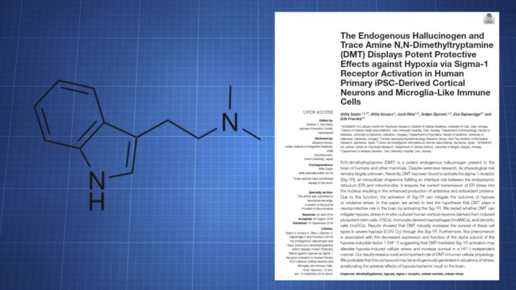
This paper, which was published on September 14, 2016, examines the effect of N,N-Dimethyltryptamine (DMT) on hypoxia-induced cellular changes. It specifically looks at the role of the Sigma-1 receptor.
Abstract: DMT is capable of reducing cell death when cells are placed in a hypoxic environment. Sigma-1 receptor activity is involved in that effect.
Authors: Attila Szabo, Attila Kovacs, Jordi Riba, Srdjan Djurovic, Eva Rajnavolgyi, and Ede Frecska
Link: http://journal.frontiersin.org/article/10.3389/fnins.2016.00423/full
Contents
Introduction
The Sigma-1 receptor is known to play a role in cellular responses to hypoxia and DMT is a known ligand for the receptor. DMT’s effect on Sigma-1 has been studied to some degree and this study further explores DMT’s effects to see if it can protect cells during hypoxia.
Cell Types
Here are the cell types that were used:
- Cortical neurons derived from human-induced pluripotent stem cells (iPSCs).
- Monoctye-derived macrophages (moMACs)
- Monocyte-derived dendritic cells (moDCs)
Results
Sigma-1 Expression in iPSC-Derived Neurons
Expression of Sigma-1 hadn’t been investigated in iPSC-derived cortical neurons, whereas it has been looked at in moMACs and moDACs, both of which express high levels of Sigma-1.
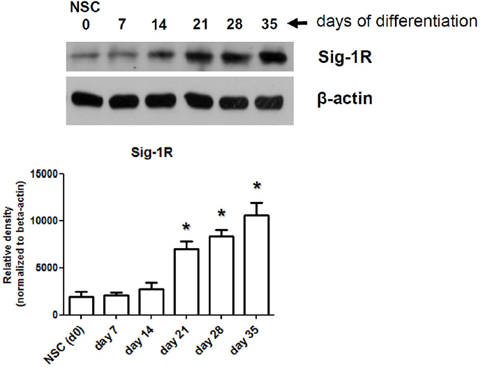
Sigma-1 protein expression in differentiating cortical neurons
The neural stem cells were found to have a low baseline expression level. But, as the cells differentiate into neurons, the expression increases, ultimately reaching a peak at the end of the differentiation.
DMT Boosts Survival During Hypoxia
To see how cells would respond during hypoxia, an environment with 0.5% oxygen was used.
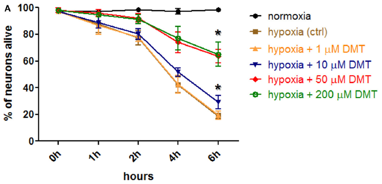
DMT’s effect on neuron survival
Cell death, more so with the neurons, was observed and it was reduced with DMT. DMT was used at a concentration ranging from 10 – 200 uM, with the efficacy depending on the cell type.
- Neurons (after 6 hours of hypoxia)
- Control = 19% survival
- 10 uM DMT = 31% survival
- 50 uM DMT = 64% survival
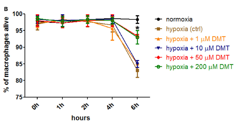
DMT’s effect on moMAC survival
Significant beneficial changes were seen with neurons beginning at 10 uM, whereas 50 uM had an effect in moMACs and moDCs. No difference in the MACs/DCs was seen between 50 uM and 200 uM. Also, the survival rate was still relatively high in moMACs and moDCs by the 6 hour mark, though the drop that was seen could be reduced with DMT.
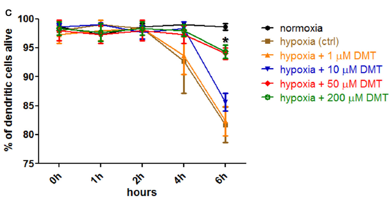
DMT’s effect on moDC survival
DMT Affects HIF-1a Expression
Hypoxia-inducible factor 1-alpha (HIF-1a) normally increases during hypoxia, a change that can largely be prevented by DMT at 50 uM.
All of the control cells showed higher levels of HIF-1a protein expression after 6 hours of hypoxia. When DMT was used, the increase was much lower.

DMT’s effect on HIF-1a expression
VEGF (vascular endothelial growth factor), a protein connected with HIF-1a, appears to increase (as seen by measuring mRNA expression) during hypoxia. This change was blunted by DMT as well.
When a non-hypoxic environment is used, DMT has no effect on HIF-1a.
Sigma-1 Is Involved In These Effects
To make sure Sigma-1 is actually involved, two separate methods were employed. First, the Sigma-1 gene was silenced. Second, an antagonist was used.
In the first test, silencing led to the following receptor downregulation:
- Over 93% in neurons
- Over 96% in moMACs
- Over 91% in moDCs
Silencing of the gene got rid of DMT’s ability to boost cell survival during hypoxia. It also blocked the HIF-1a modulation.
Sigma-1 silenced cells treated with DMT did worse in hypoxia than the control cells.
As for the antagonist experiment, BD1063 was used. It successfully blocked DMT’s effect on survival and HIF-1a.
Summary
Hypoxia is capable of causing significant cell death in iPSC-derived cortical neurons, moMACs, and moDCs. When DMT is used, a concentration of 10 uM – 50 uM can improve cell survival in neurons, while 50 uM improves survival in the other cells.
Just as cell death is induced, HIF-1a is normally increased along with VEGF mRNA expression. DMT also greatly reduces these effects.
Conclusion
DMT appears to exert a protective effect during hypoxia and those effects are connected to Sigma-1 receptor agonism.